
2D radiology program
AIM:
Introduce the participants to a systematic approach of the 2D diagnostic imaging aspects.
OBJECTIVES:
Discuss the X-Ray techniques (analog, digital):
Intra oral radiology.
Extra oral radiology.
Be aware of the errors of Intra-Oral Radiology
Be aware of the errors of Extra-Oral Radiology
Review the 2D radiographical anatomical landmarks (Maxilla and Mandible)
Discuss the inclusion of prosthetic considerations in the complete treatment plan
Careful assessment of available hard and soft tissues.
Diagnosis and planning of surgical procedures
Assess the potential risks and complications to surgical procedures in variances to the average radiographic appearance
AT THE END OF THE COURSE, THE PARTICIPANTS WILL BE ABLE TO:
Successfully take 2D imaging
Distinguish normal from abnormal radiographic appearances.
Develop a differential diagnosis of the radiographic findings
Use 2D imaging to formulate a proper diagnosis
Use diagnostic adjuncts to clinically evaluate and overcome shortcomings in various imaging techniques
2. 3D Radiology Program
AIM:
Introduce the participants to the value and limitations of various 3D imaging techniques in the dentist’s daily work.
OBJECTIVES:
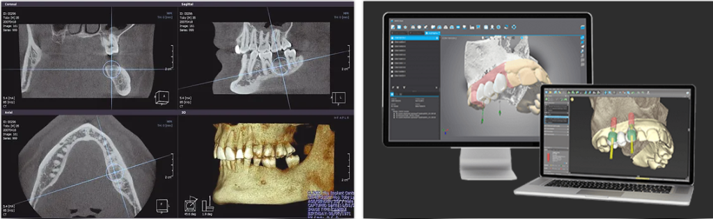
Discuss the 3D imaging techniques
Computed tomography (CT)
Cone beam computed tomography (CBCT)
Discuss the advantages and limitations of the 3D imaging techniques (CT, CBCT).
Review the 3D radiographical anatomical landmarks (Maxilla and Mandible)
Discuss the use and the manipulation of various 3D software.
Introduction to the reading of three-dimensional radiological records.
Introduction to the guided surgery
Fabrication and utilization of the radiographic guide
Transformation of the radiographic guide into a directional surgical guide
Fabrication of the full surgical guide
Introduction to the pre- and postoperative 3D diagnosis in:
Dental implantology
Periodontology
Endodontics
AT THE END OF THE COURSE, THE PARTICIPANTS WILL BE ABLE TO:
Successfully manipulated 3D imaging software
Successfully read and interpretation 3D imaging records
Distinguish normal from abnormal radiographic appearances.
Develop a differential diagnosis of the radiographic findings
Use 3D imaging to formulate a proper diagnosis
Use 3D imaging to realize a surgical guide
- Course Duration (Days): 2 Days
- Course starting on : Registration is open to be held in Q4. 2024
- Total Points: 20 CE
- Fees: 1,290 CAD
- Course delivery: Online